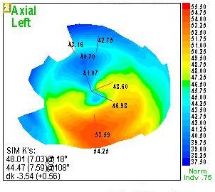

Several surgical procedures have been used in an attempt to improve visual acuity when spectacles and contact lenses do not provide adequate vision correction. The vast majority of PMD patients are managed using spectacles and contact lenses. In rare cases, patients may present with a sudden loss of vision and excruciating ocular pain due to corneal hydrops or spontaneous perforation. Visual signs and symptoms include longstanding reduced visual acuity or increasing against-the-rule irregular astigmatism leading to a slow reduction in visual acuity. Unless corneal topography is evaluated, early forms of PMD may often be undetected however, in the later stages PMD can often be misdiagnosed as keratoconus.

Ocular signs and symptoms of patients with PMD differ depending on the severity of the condition. The prevalence and aetiology of this disorder remain unknown. The condition is most commonly found in males and usually appears between the 2nd and 5th decades of life affecting all ethnicities. Pellucid marginal corneal degeneration (PMD) is a rare ectatic disorder which typically affects the inferior peripheral cornea in a crescentic fashion.


 0 kommentar(er)
